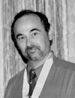
The University Record, September 27, 1999 By Wanda Monroe
Office of the Chief Information Officer

Lloyd Stoolman was attempting to help his students learn more about the properties of diseased tissues when he came up with the idea of using the Web to simulate the experience students normally have peering through the microscope in a pathology laboratory. His idea led to the development of Web-based courseware called “The Virtual Microscope.” The courseware is comprised of two innovative applications that allow students to study the microscopic structures of diseased organs, tissues and cells—anywhere, anytime from their networked computers.
“The Virtual Microscope” provides access to teaching materials previously available only in laboratories, and uses interactive tools to simulate the laboratory experience.
The application includes hundreds of high resolution, true-color digital images and interactive exercises that help students recognize the macroscopic and microscopic hallmarks of disease processes in general pathology and hematopathology. “The structural changes that occur in organs and tissues ravaged by disease helps students understand the impact diseases have on normal function,” says Stoolman, associate professor of pathology.
Each exercise includes multiple images of the tissue or organ along with descriptions and study questions. The commercial image server software powering the application allows students to magnify and pan the online digital “slides,” simulating the experience of a laboratory microscope. The program also provides instant feedback on the study questions included in the exercises.
Thanks to the interactive tools available in the Web environment students can design exercises that stimulate active learning, supplementing the traditional lecture and laboratory formats. Because the images are available online 24 hours a day, seven days per week, students can learn at their own pace. Some students use the materials to prepare for class, others use it exclusively for review, and some do both. However it is used, it benefits students by giving them more control over and responsibility for the educational process, Stoolman says.
Stoolman notes that many colleagues in the Medical School and the Information Technology Division contributed to the project. “Dr. Gerald Abrams’ exquisite photomicrographs were critical to the success of Cyberscope631, one of the two Virtual Microscope Web sites currently in operation.” He also cites Michael Lougee, Douglas Gibbs, Thomas Peterson, Casey White, Dino Anastasia, Joseph Fantone, Bruce Friedman and Richard Lieberman as “just a few who helped ensure that ‘The Virtual Microscope’ became a reality.”
“This project provides new challenges every year,” Stoolman says. “Updating and improving the content of ‘The Virtual Microscope,’ optimizing its performance and assuring its compatibility with the myriad of workstations used by our students is a dynamic and ongoing process. This project has deepened my respect for the computer experts and administrators who keep the computing infrastructure humming at the University in general and at the Medical School in particular.”
“The Virtual Microscope” was one of five finalists in the Education and Academia category of the 1999 Computerworld Smithsonian Program. This category was the largest in the competition, drawing 13 entries from the U-M and 75 overall.
Scott McNealy, chairman and chief executive officer of Sun Microsystems Inc., nominated “The Virtual Microscope” and 12 other computer applications from the U-M for the Computerworld Smithsonian Award Program. A total of 14 U-M entries—the 14th was the M-Pathways Project, nominated by Peoplesoft—were selected for inclusion in the 10th anniversary collection of computer applications added last spring to the Permanent Research Collection on Information Technology at the Smithsonian’s National Museum of American History.
“The fact that our highly focused and somewhat obscure application was selected as one of the five best in such a diverse field indicates that the novel application of Web-based technologies used in The Virtual Microscope resonated with educators in many disciplines,” Stoolman says. “The content developers and technical specialists who made it happen, as well as the administrators who nurtured its development, can be proud of the accomplishment.”
“The Virtual Microscope is an innovative example of support for the learning environment we are seeking to develop here at the University,” said Jose-Marie Griffiths, University chief information officer. “It is also an example of a University-based resource that has a potential value for a much larger community.”
The Virtual Microscope currently has two Web sites, one that profiles general and organ systems pathology for dental and graduate students, http://141.214.6.12/cyberscope631/, and one that focuses on hematopathology for medical students, http://141.214.6.12/virtualheme99.

