More wrinkles found on ‘protected’ skin of smokers
In classic movies cigarette smoking was used as shorthand to convey sultriness and beauty. In the real world the connection between smoking and one’s appearance has more to do with premature signs of aging and less to do with glamour and refinement.

A new study from the U-M Health System adds another dimension to the link between cigarette smoking and skin damage—smoking may be associated with a higher degree of aging on areas of skin, such as the inside of the upper arm, that normally are not exposed to sunlight.
“We examined non-facial skin that was protected from the sun and found that the total number of packs of cigarettes smoked per day and the total years a person has smoked were linked with the amount of skin damage a person experienced,” says Dr. Yolanda Helfrich, lead author and assistant professor of dermatology at the Medical School.
The researchers developed a photonumeric scale that easily can be reproduced by other medical institutions to measure the degree of aging on patients’ skin. The nine-point scale used information from photographs of the inside-upper-arm skin of the 77 participants.
Two medical residents and a medical student were asked to look at the photographs and assign a grade in which zero represented no fine wrinkling and eight represented severe fine wrinkling. The same three people reviewed photos of the participants one year later, and the scores were used to determine the level of increase in the skin damage.
Researchers also collected data from interviews, such as age, ethnicity, history of cigarette smoking, use of non-steroidal anti-inflammatory drugs, use of dietary or herbal supplements, sun exposure, sunscreen use, tanning bed use and, for women, how many children they had given birth to, hormone therapy use and oral contraceptive use.
In all, among participants who were 45 years or older, the degree of skin aging was found to be significantly higher in smokers than nonsmokers.
In addition to Helfrich, authors were Dr. Abena Ofori, Ted Hamilton, Jennifer Lambert, Anya King, Dr. John J. Voorhees and senior author Dr. Sewon Kang, all of the Department of Dermatology, and Dr. Le Yu, now with Cedars Sinai Medical Center in Los Angeles.
The study appears in the March issue of the journal Archives of Dermatology.
—Katie Gazella, Medical School Public Relations
Forced rankings of employees bad for business
Ranking systems that require managers to evaluate the performance of an employee against other employees can hurt productivity, says a researcher at the Ross School of Business.
“Individuals will care less about performing better on a given task and will, instead, shift their focus to performing relatively better on a scale comparison—in other words, being surpassed in rank,” says Stephen Garcia, adjunct assistant professor of management and organizations at the Ross School and assistant professor at the Ford School of Public Policy.
General Electric is the most famous proponent of the forced-ranking model, but other companies have used it in some form at one time or another, including Microsoft, Pepsi, Goodyear, Ford and Capital One.
In a new study in the current issue of Organizational Behavior and Human Decision Processes, Garcia and colleague Avishalom Tor of the University of Haifa examine the forced-ranking system by conducting a series of studies of task versus scale comparisons.
Comparisons concern relative outcomes or standing in specific tasks (e.g., anticipated earnings of one’s company versus a partner company in a joint venture), while comparisons on the scale—the metric that defines a standard—occur at a more general level (e.g., those concerning a company’s standing on annual earnings).
The researchers found that rivals ranked near the top of a standard are more competitive and less cooperative than those far from a standard. Only 25 percent of the study participants were willing to maximize joint gains when they and their rivals were ranked Nos. 1 and 2, compared to 79 percent when ranked Nos. 101 and 102. When the rivals’ relative standing on the scale was not in jeopardy, however, participants uniformly behaved more cooperatively—74 percent for Nos. 1 and 2 and 77 percent for Nos. 101 and 102.
Similar results were found in a second study of the Fortune 500. Competitive behavior not only was more prevalent among the highly ranked, but also among those at the bottom vying to make it in to the Fortune 500.
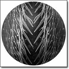
Artists engage nature in a micro/macro world
The growing connections between art and science are the focus of The Seduction of Scale, a group exhibition at the Life Sciences Institute Library.
As recent developments in scientific research have provided the artist/designer with important new tools, materials, methodologies and issues, art and design have illuminated many social issues related to current scientific practices for the scientist, biologist and geneticist.
The Seduction of Scale, available for viewing 11 a.m.-5 p.m. weekdays now through April 30, reveals how art making can merge art, science and imaging technologies to explore those fundamental issues that both artists and scientists must address as they employ and interpret new advances in scientific research.
Works include those by Beverly Fishman, David Mann, and three professors from the School of Art & Design: Jan-Henrik Andersen, Patricia Olynyk and Brad Smith. “This exhibit represents a microcosm of the vast array of science-inspired art that evokes what nature reveals and scientists and artists alike explore,” Olynyk says.
The Seduction of Scale is a part of a larger partnership among the Life Sciences Institute, the School of Art & Design, and the Museum of Art to actively explore the intersections of art, science and technology. This program also has assembled a collection of works now on permanent display in the LSI, and initiated its first joint faculty appointment with the School of Art & Design in Olynyk, an associate professor at SOAD and research associate professor at LSI.
—Kate West, SOAD and Robin Stephenson, LSI
Lab-grown heart muscle promising, not patient-ready
Some day heart attack survivors could have a patch of laboratory-grown muscle to replace areas that died during the attack. Children with defective valves could get new ones that grow with them, rather than being replaced every few years. And people with clogged or weak blood vessels may get a natural replacement, instead of a factory-made one.
These possibilities all are within reach and could transform heart care, say Medical School researchers in the new issue of the journal Regenerative Medicine.
Technology has advanced so much in recent years, they write, that scientists are closer than ever to bioengineering entire areas of the heart, as well as valves and major blood vessels. But hurdles still remain before the products of this tissue engineering are ready to be implanted in patients, the team notes. Among the obstacles: determining which types of cells hold the most potential, and finding the best way to grow those cells to form viable cardiac tissue that is strong, long lasting and structured at a cellular level like natural tissue. The article reviews the current state of cardiac tissue engineering at the U-M Cardiac Surgery Artificial Heart Laboratory and worldwide.
“Although there remain tremendous technological challenges, we are now at a point where we can engineer first-generation prototypes of all cardiovascular structures: heart muscle, tri-leaflet valves, blood vessels, cell-based cardiac pumps and tissue engineered ventricles,” says Ravi Birla, senior author and director of the Artificial Heart Laboratory.
In December, Birla and Yen-Chih Huang published a paper describing their success in growing pulsing, three-dimensional patches of bioengineered heart muscle, or BEHM. Previously, the group described the results of a self-organization strategy, showing that it was possible to engineer heart muscle that closely resembles normal physiology without synthetic scaffolding material.
The new article by Birla and lead author Louise Hecker, a graduate student in the Department of Cell & Developmental Biology, describes the bioreactor that the team uses to grow BEHM and details many other discoveries that have been made as well as hurdles that lie ahead.
—Kara Gavin, UMHS Public Relations