At a new state-of-the art electron microscopy lab within the Life Sciences Institute, Georgios Skiniotis “quick-freezes” protein complexes inside a thin, icy shell before viewing them, to preserve their natural structure.
He plunges the samples into liquid ethane at minus 256 degrees Fahrenheit. The freezing occurs so fast that the resulting ice shell is vitreous, meaning it has glass-like properties and contains no crystals that could damage the specimen.
The freezing process is part of an emerging imaging technique called electron cryomicroscopy, or cryo-EM for short. Structural biologist Skiniotis is an expert in the field, and he designed and supervised the construction of the $8 million LSI Cryo-EM Facility.
The three-microscope suite was completed this spring and allows U-M structural biologists to visualize biological specimens at the molecular scale. It is one of a dozen or so dedicated electron cryomicroscopy labs in the country, says Skiniotis, a research assistant professor who joined LSI in October 2008.
“Cryo-EM is an extremely powerful tool for studying the structure of protein complexes,” Skiniotis says. “It enables us to visualize in detail macromolecular architectures that are well-preserved within the vitreous ice layer, at conditions close to physiological.”
A transmission electron microscope uses an electron beam that is transmitted through the thin sample under study. The interaction of the electron beam with the sample results in the formation of an image, which subsequently is magnified and focused by a series of electro-magnetic lenses, and finally recorded on a photographic plate or a CCD camera.
Several thousand views of the specimen, observed from various angles, then are combined by a computer to reconstruct a 3-D model of the sample’s structure.
“These technologies allow us to figure out how proteins come together, like pieces of a 3-D puzzle, and form complex macromolecular architectures that perform different types of work according to their structure,” Skiniotis says.
The lab was built in the basement of the Life Sciences Institute to minimize vibrations that can degrade image quality. Temperature, humidity and airflow fluctuations can affect specimen preparation and image quality, so environmental controls in the seven-room suite are stringent. The temperature is maintained at 67 degrees, the humidity level is 30 percent and airflow in the rooms is limited to less than 2 feet per minute.
“These instruments are very sensitive to airflow, and the slightest movement can blur an image,” Skiniotis says. “We want these images to be as crisp as possible. Every detail of the facility’s layout has been designed so that these instruments will perform optimally.”
The lab has 100-, 120- and 200-kilovolt transmission electron microscopes, and a 300-kilovolt instrument also is planned. Skiniotis and his lab colleagues usually visualize samples at a magnification of about 50,000 times, and the resolution of the resulting 3-D models is typically around 10-20 angstroms.
“The cryo-EM lab is our newest effort to bring scientists together to tackle the tough but crucial biological questions,” says Alan Saltiel, the Mary Sue Coleman Director of LSI. “Now that our building is full of great faculty laboratories, it is critical to add the tools that can take the science to the next level.”
The new facility was funded by the Office of the President, the Office of the Executive Vice President for Medical Affairs, the Office of the Provost and Executive Vice President for Academic Affairs, the Endowment for the Basic Sciences at the Medical School, and LSI.
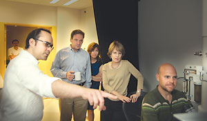
Coleman, a biochemist, toured the lab recently and says she came away impressed by what she’d seen.
“To see this project move so quickly from concept to completion is very gratifying,” Coleman says. “This is an excellent example of how we’re making strategic investments across campus to advance our ability to do the very best science.”
Structural biologists at LSI will use the new microscopes, and Skiniotis says he’s already formed collaborations with researchers in the U-M chemistry, biological chemistry and pharmacology departments.