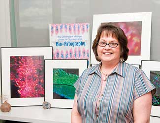
Those neon sea creatures are actually dying photoreceptors in the human retina. And that intense blue-green hot air balloon-looking thing, complete with gondola, is really a salamander sperm cell.

These images are among those collected through the nearly matchless BioArtography project, which celebrates microscopic tissue photography as art. The images are organized by Rebecca Pintar from a second-floor office overlooking a window-lit atrium in the Biomedical Science Research Building.
BioArtography is a product of The Center for Organogenesis, which unites scientists from many fields to study organ formation, function and disease. The goal of their research is to use new information to design effective strategies to treat disease and repair damaged organs.
Director Deborah Gumucio and member Sue O’Shea founded BioArtography in 2005 as a fundraising venture for training activities in the center. “Every day, they were seeing beautiful images in the microscope and they believed that others might also be struck by this beauty,” says Pintar, administrative specialist at the center.
BioArtography generates its highest public profile during the Ann Arbor Art Fairs, July 16-19, as the most striking images are sold from a booth on South University across from the Brown Jug. Pintar says booth sales raise more than $10,000 each year.
All monies generated from BioArt sales — minus 5 percent which goes to the submitting artists — fund special projects of graduate students and pre- and post-doc fellows associated with the center, says Pintar, who has worked at U-M for nearly 29 years. In addition, each piece of BioArt comes with an explanation of the science, written in lay terms.
While stains typically used in micrograph photography add color, Pintar often will use Photoshop to sharpen images and intensify colors. The rich yellows, blues and reds in one photo of a basil cell skin cancer have drawn comparisons to a Van Gogh painting.

To select new images to be reproduced and sold at the art fairs, the center asks a three-member committee typically made up of representatives from University arts related groups, including the School of Art & Design and Arts at Michigan, to choose photos. Their choices are presented to a BioArtography committee and approximately 25 new images are chosen to add to the best selling photos from previous years.
Pintar coordinates the booth display, manages image inventory and schedules the more than 40 people who work the booth every year.
“It is really a wonderful experience for all to interact with the public and answer questions about the images,” she says. Her primary job during the art fairs is to keep the booth stocked and she constantly is printing, matting, and framing the works of art, sometimes up to 12 hours a day.
The work has been displayed at the Ann Arbor Hands-on-Museum, the University Hospital Gifts of Art and the Michigan Life Sciences Expo in Lansing, and it can be found throughout the BSRB where the center’s offices/labs are located. A future display is planned at the Exhibit Museum of Natural History. Images also can be ordered online at www.bioartography.com.
After working briefly in 1975 as a typist in the Department of Dermatology, Pintar, an Ypsilanti native, left the University. She worked at Ford Motor Co., then returned to U-M in 1980 where she worked in the Department of Radiology, School of Public Health and the Institute of Gerontology. She took her position with The Center for Organogenesis in 2000.
Pintar serves as the center’s primary administrator with tasks that include financial management, a seminar series, a graduate course and an International Symposium on Organogenesis, which typically draws more than 250 attendees.
When Pintar isn’t working, she enjoys gardening, camping, cooking and spending time with husband David, who retired in 2004 from Ford. They have a Chinese pug named Curly.
The weekly Spotlight features staff members at the University. To nominate a candidate, please contact the Record staff at [email protected].

